
2015 IMR Case of the year
Clinicians from across the globe took part in the 2015 Case of the Year Contest to show the outstanding results they're accomplishing using Philips IMR (Iterative Model Reconstruction). Cases* were reviewed by a panel of medical professionals. See our “Meet the judges” area. IMR is an image reconstruction solution that has set a new direction in CT image quality with industry-leading low-contrast resolution and virtually noise-free images. Innovations in hardware and the reconstruction algorithm result in a reconstruction speed – less than three minutes for the majority of reference protocols – that allows model-based benefits to be achieved in even the most demanding applications.
Images created with:
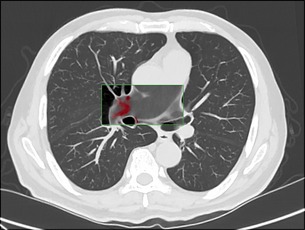
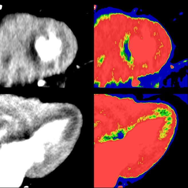
Winner: Non-contrast Pulmonary Embolism
Einstein Healthcare Network United States Dr. Terence Matalon, Dr. Thomas Reilly, & Team “On a non-contrast CT chest, IMR allowed for improved visualization of hyper-dense thrombus in the pulmonary artery (pulmonary embolism). This was confirmed with subsequent contrast enhanced CT.”
Parameters: kVp: 120 mAs: 50 Scan length: 27.5 cm Scan time: 2.2 sec CTDIvol: 3.3 mGy DLP: 90.7 (mGy*cm) Scanner: iCT Dose: 1.7 mSv* *AAPM Technical Report 96
Our finalists
Thank you to all those that took the time to submit their IMR cases* to show the world what they and Philips IMR are accomplishing in CT. We know that dose, image quality and diagnostic confidence matter in today’s challenging healthcare environment like never before, and we were excited to receive so many great cases illustrating the difference IMR can make in meeting these challenges. Congratulations to the following finalists. Your work helps show the difference virtually noise-free images**, industry-leading low-contrast resolution, and low-dose scanning can make to impacting patient care.
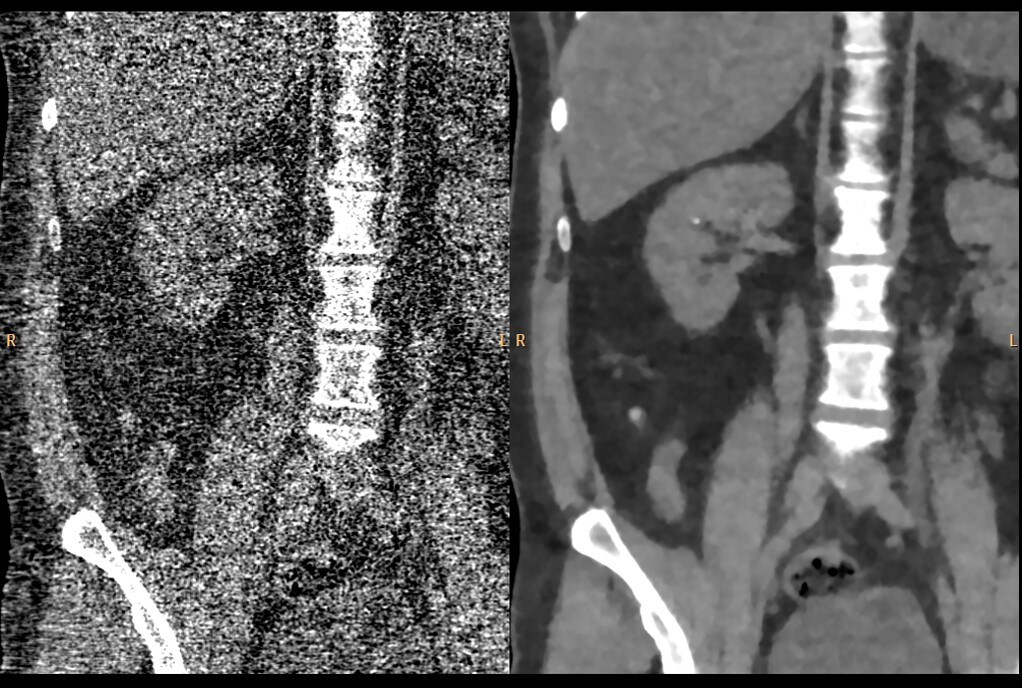
NCCT KUB
Post Graduate Institute of Medical Education and Research (PGIMER) “This is an exceptional IMR case as NCCT KUB was done at a radiation dose exposure (0.6 mSv) equivalent to Xray KUB and yet produced images with exquisite details. A tiny 3 mm calculus was seen in right kidney on IMR images which was not seen on other imaging modalities (USG, KUB) as well as on images produced by standard reconstruction. Hence low dose CT with IMR benefited this case to a great extent.”
India
Dr. Khandelwal & Team
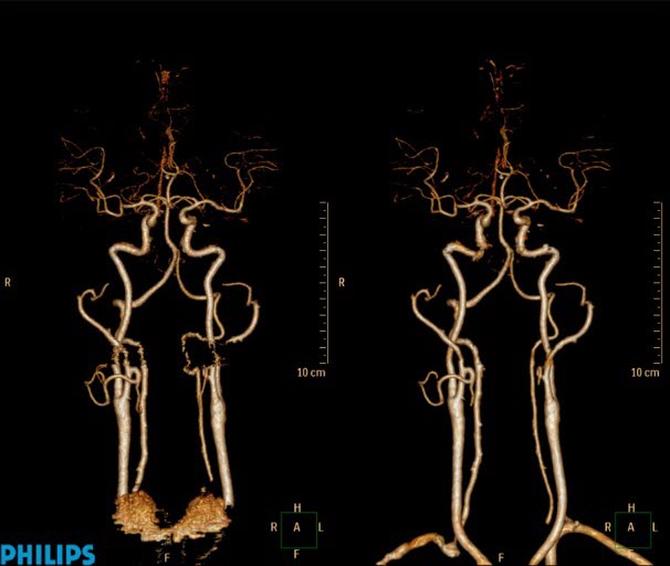
Head & neck angiogram
Yonsei University College of Medicine - Severance Hospital “Comparing FBP and IMR, especially in the shoulder area, you will see the removal of streak artifact. O-MAR with IMR can reduce dental artifact.”
South Korea
Dr. Ho Jun Lee & Team
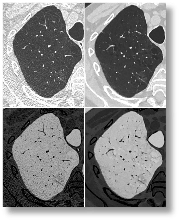
Emphysema / ground glass nodule
Universitair Ziekenhuis Brussel “This scan with only half the dose as used in daily practice and reconstructed with IMR depicted the extent of the emphysema more clearly. The ground glass nodule showed a sharper delineation relative to the surrounding lung parenchyma on the second scan with half dose compared to the scan with full dose.”
Belgium
Dr. Brussard & Team
Triple rule out
Chungang University Hospital “PCTA (prospective coronary CTA) showed significantly improved subjective and objective image quality parameters for coronary vessels with IMR compared to FBP and iDose⁴. PCTA using 80KVp and IMR could be applicable in routine clinical setting with less radiation dose.”
South Korea
Dr. Jae Suung Seo, Dr. Sung Bin Park & Team
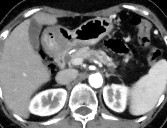
Pancreatic cancer
Seoul National University Hospital “Pancreatic cancer on Chemotherapy (3 times f-u scanning). Reduced lesion size after CTx, better visibility in small lesion with IMR than FBP and iDose⁴. ”
South Korea
Dr. Jeong Min Lee & Team
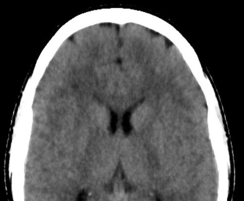
Ischaemic white matter disease, IMR vs MRI
Sydney Xray Bondi Junction “IMR was capable of demonstrating the multiple lesions in the brain that was confirmed on the MRI scan. Nice improvement in image quality and compares well with MR.”
Australia
Dr. Sesel & Team
Myocardial perfusion
Ehime University School of Medicine “Due to detection of myocardial ischemic area in preoperative evaluation, Perfusion CT with IMR is able to detect it. We think that it might be possible to diagnosis with high accuracy, because Cardiac CT is able to assess morphology and function simultaneously. IMR can be used to conduct dynamic perfusion study with low radiation dose. It is possible to enhance contrast between ischemic and normal myocardium by using low-kV technique with IMR.”
Japan
Dr. Mochizuki, Teruhito Kido & Hikaru Nishiyama
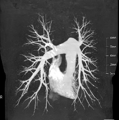
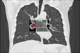
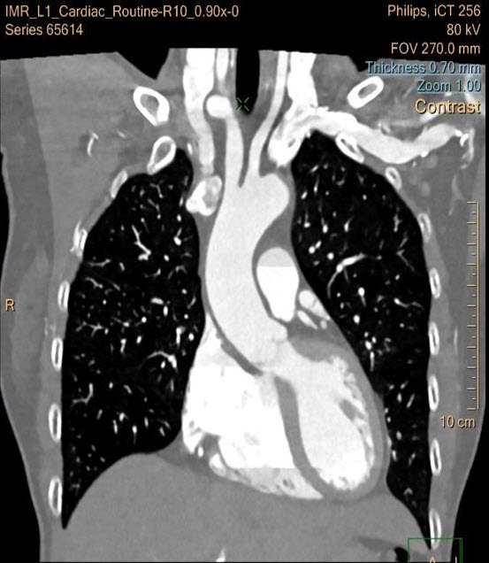
Pulmonary artery CTA
Hokkaido University Hospital “IMR images are very effective to understand the orientation of pulmonary vessels will be used during an operation. We achieved lower dose than the radiation dose of routine dose chest CT.”
Japan
Tsukasa Sasaki & Team
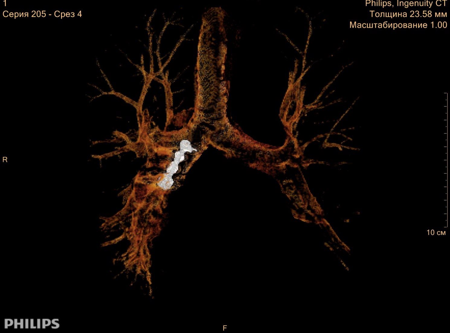
Aspiration of teeth
Federal Almazov North-West Medical Research Centre “Nice imaging of artificial teeth in the bronchus. With IMR we could create very demonstrative 3D model”
Russia
Dr. Gleb & Team
Emmanuel Coche, MD, PhD Head of Department of Radiology Cliniques Universitaires St-Luc Brussels, Belgium
Claudio Smuclovisky, MD, FACC, FSCCT Director SFMI Cardiovascular Institute Holy Cross Hospital Fort Lauderdale, Florida
Kenneth Lau, MBBS, FRACR Associate Professor Head of CT and Thoracic Imaging Monash Health Melbourne, Victoria, Australia
* Results from case studies are not predictive of results in other cases. Results in other cases may vary. ** Image noise as defined by IEC standard 61223-3-5. Image noise was assessed using reference body protocol, on a phantom. Data on file.